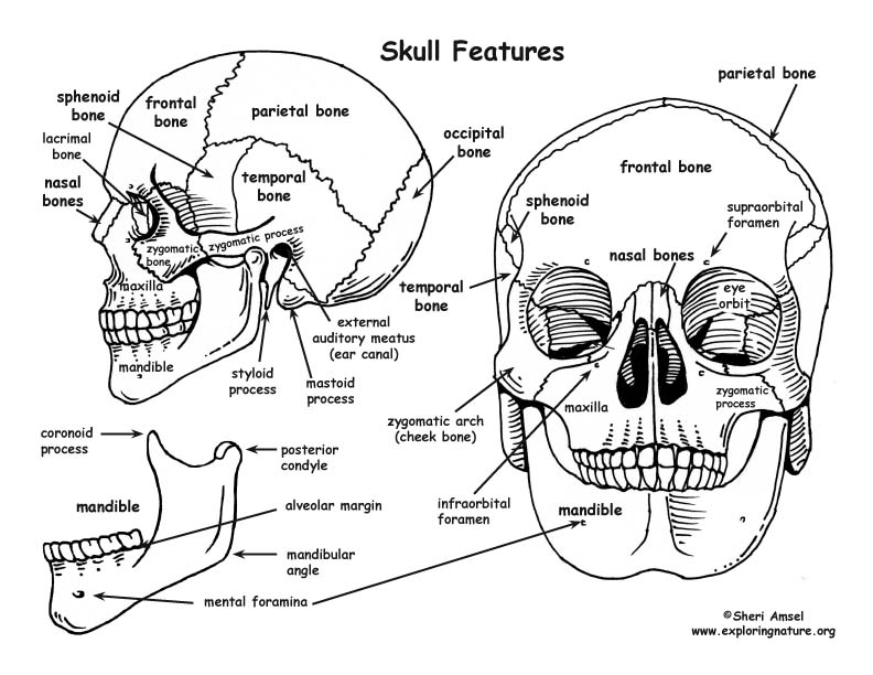
The skull is made up of the cranial bones (of the cranium) and the facial bones, which include the mandible. The joints of the cranial and facial bones are called sutures.
The cranium surrounds and protects the brain and the organs of hearing and balance. The facial bones form the structure of the face, hold the eyes, and the organs for taste and smell and anchor the teeth. They have the openings for air and food. The whole skull anchors muscles that hold the head up, allow us to chew, and form facial expressions. Most of the bones of the skull are flat bones, except the mandible. The mandible is attached to the skull by a type of hinge joint. It is the biggest, strongest bone of the skull.
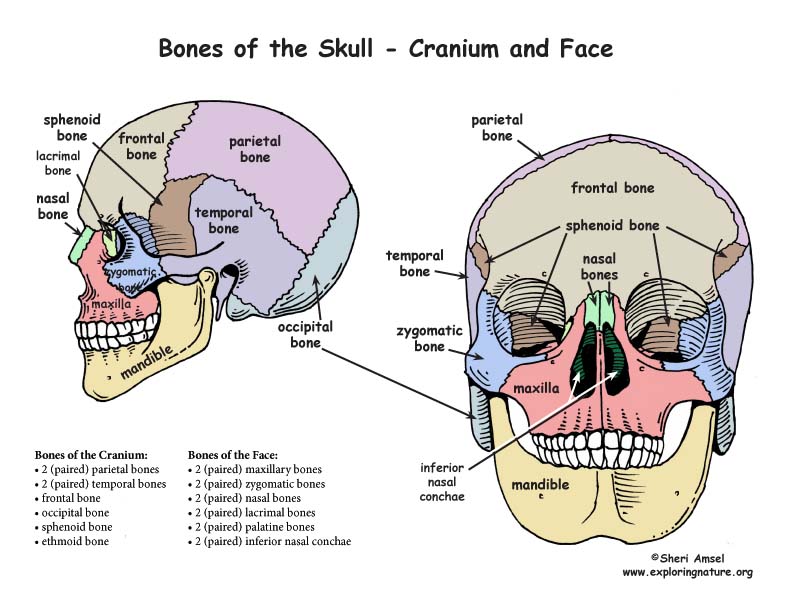
The cranium is made up of 8 bones:
• 2 (paired) parietal bones
• 2 (paired) temporal bones
• frontal bone
• occipital bone
• sphenoid bone
• ethmoid bone
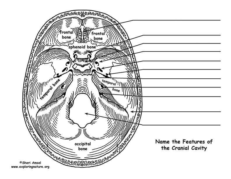
The frontal bone is located on the anterior cranium and includes the following features:
1) It makes up the roof of the eye orbits.
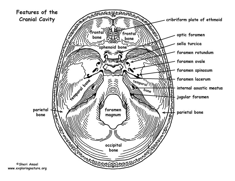
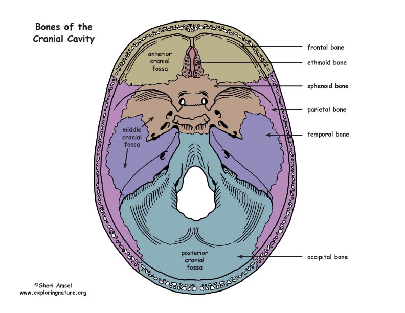
2) Inside the skull, it forms the anterior cranial fossa, which contains the frontal lobes of the cerebrum.
3) It has the supraorbital foramen, where the supraorbital artery and nerve pass out of the skull onto the forehead.
4) It contains the frontal sinus (part of the paranasal sinuses).
5) It makes up (with the parietal bone) of the anterior fontanelle.
The paired parietal bones make up the top and lateral aspects of the cranium.
The occipital bone is located on the back of the cranium and includes the following features:
1) Internally, it forms the posterior cranial fossa where the occipital lobe of the brain sits.
2) It contains the foramen magnum – the passageway between the vertebral column and the cranial cavity.
3) It has the external occipital protuberance.
The paired temporal bones are located on either side of the cranium, inferior to the parietal bones and contain the following features:
1) They form the zygomatic process (posterior portion of the zygomatic arch).
2) They form the mandibular fossa (underneath the zygomatic process where the jaw articulates with the cranium via the posterior condyle). This is the temporomandibular joint – TMJ.
3) They contain the external auditory meatus (external ear canal)
4) They form the styloid process (a muscle attachment site)
5) They form the mastoid process (the lump behind the ears where neck muscles are anchored).
6) They contain the stylomastoid foramen (between the styloid and mastoid processes, where the facial nerves leave the skull)
7) They form the posterior part of the middle cranial fossa (where the temporal lobes of the brain sit).
8) They contain the jugular foramen (the most posterior foramen, where the internal jugular vein brain the brain).
9) They contain the carotid canal (an opening where the internal carotid artery enters. It passes so close to the inner ear, you can sometimes hear the blood thundering past).
10) They contain the foramen lacerum (a jagged opening allowing some nerve passage).
11) They contain the internal acoustic meatus (in the medial cranial fossa – which contains the internal auditory artery, facial nerve and the vestibulocochlear nerve).
The sphenoid bone contains the following features:
1) It forms the paired sphenoid sinuses.
2) It forms the sella turcica (where the pituitary gland sits).
3) It forms the anterior portion of the middle cranial fossa.
4) It has the optic foramina (where the optic nerve and ophthalmic artery pass).
5) It has the superior orbital fissure (where the cranial nerve to the eye muscles pass).
6) It has the foramen rotundum (small round hole).
7) It has the foramen ovale (large oval hole).
8) It has the foramen spinosum (small hole).
The ethmoid bone is located between the nasal bones of the face and has the following features:
1) It forms the crista galli on top (superiorly) – the “cocks comb.”
2) It forms the cribriform plate (a horizontal plate beneath the crsita galli). It is full of holes (olfactory foramina) where the olfactory nerves pass to pick up smell.
3) It forms the perpendicular plate (which projects doen to be part of the nasal septum).
4) It has the ethmoid sinuses (part of the paranasal sinuses)
5) It forms the superior and middle nasal conchae (extending laterally and inferiorly)
The face is made up of 14 bones:
• mandible
• vomer
• 2 (paired) maxillary bones
• 2 (paired) zygomatic bones
• 2 (paired) nasal bones
• 2 (paired) lacrimal bones
• 2 (paired) palatine bones
• 2 (paired) inferior conchae
The mandible (lower jaw) is the largest strongest bone of the face and contains the following features:
1) It has the mandibular angle (angle of the jaw below the ear).
2) It has the coronoid process (anterior process which is the attachment site for the temporalis muscle which elevates the jaw during chewing).
3) It has the mandibular condyle (the posterior process that is part of the TMJ and articulates with the mandibular fossa on the zygomatic process of the temporal bone).
4) It has the mandibular notch (between the two processes).
5) It forms the alveolar margin (where the lower teeth are inserted).
6) It has the mandibular foramina (inside the ramus of the mandible) there the nerves to the jaw pass.
7) It has the mental foramina (on the anterior and lateral (outside) mandible) where the blood vessels and nerves to the chin and upper lip pass.
The paired maxillary bones are fused medially and contain the following features:
1) They make up the upper jaw and central face.
2) They form the alveolar margin (where the upper teeth are inserted).
3) They include the palatine processes (the hard palate) that projects posteriorly.
4) They include the frontal processes (medial and inferior eye orbit by the bridge of the nose).
5) The form the maxillary sinuses (the largest part of the paranasal sinuses between the orbits and the upper teeth).
6) They form the zygomatic processes (the anterior part of the zygomatic arch).
7) They have the inferior orbital fissure (inside the orbit where the vagal nerve and zygomatic nerve and blood vessels pass).
8) They include the infraorbital foramen (where the infraorbital nerve and arteries to the face run).
The paired zygomatic bones (malar bones or cheek bones) sit between the zygomatic process of the maxilla and the zygomatic process of the temporal bone.
The paired nasal bones make up the bridge of the nose.
The paired lacrimal bones are made up of the medial walls of each orbit with a lateral “sulcus” through which tears can drain into the nasal cavities (which is why you get a runny nose when your eyes tear).
The paired palatine bones are inside the skull made up of the horizontal plate (which is the posterior portion of the hard palate) and the posterior, lateral wall of the nasal cavity.
The vomer is the plow-shaped bone within the nasal cavity that makes up part of the nasal septum.
The inferior nasal conchae form the lateral walls of the nasal cavity below the middle conchae of the ethmoid bone.
Assessment: Labeling the Parts of the Skull
LS1.A: Structure and Function
• All living things are made up of cells, which is the smallest unit that can be said to be alive. An organism may consist of one single cell (unicellular) or many different numbers and types of cells (multicellular). (MS-LS1-1)
• Within cells, special structures are responsible for particular functions, and the cell membrane forms the boundary that controls what enters and leaves the cell. (MS-LS1-2)
• In multicellular organisms, the body is a system of multiple interacting subsystems. These subsystems are groups of cells that work together to form tissues and organs that are specialized for particular body functions. (MS-LS1-3)