
Objectives: Observe plant cells
Materials per student (or team):
• onion
• microscope
• clean microscope slide and cover slip
• tweezers
• eye dropper
• stain - methylene blue staining solution (iodine solution can be used as an alternative stain)
• paper towel
Procedures:
1. Use the eye dropper to put a drop of water on the slide (will help to flatten out onion tissue).
2. With the tweezers, carefully peel the tissue thin sheet of cells lining the inside of one of the onion layers.
3. Lay the onion tissue gently on the slide (without wrinkling it).
4. Use the eye dropper to put a drop of stain on the onion tissue. (Be careful as it will stain skin/clothing.)
5. With tweezers, carefully place cover slip over the onion tissue (gently tap it to remove any air bubbles).
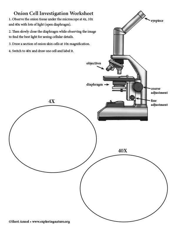
5. Observe the onion tissue under the microscope at 4x, 10x and 40x with lots of light (open diaphragm). Then slowly close the diaphragm while observing the image to find the best light for seeing cellular details.
6. Draw a section of onion skin cells at 10x magnification. Then switch to 40x and draw one cell and label it.
Study a typical plant cell to compare to your onion skin cell.
Compare to an animal cell investigation.
When you research information you must cite the reference. Citing for websites is different from citing from books, magazines and periodicals. The style of citing shown here is from the MLA Style Citations (Modern Language Association).
When citing a WEBSITE the general format is as follows.
Author Last Name, First Name(s). "Title: Subtitle of Part of Web Page, if appropriate." Title: Subtitle: Section of Page if appropriate. Sponsoring/Publishing Agency, If Given. Additional significant descriptive information. Date of Electronic Publication or other Date, such as Last Updated. Day Month Year of access < URL >.
Amsel, Sheri. "Onion Skin Cells - Investigation" Exploring Nature Educational Resource ©2005-2024. December 13, 2024
< http://www.exploringnature.org/db/view/Onion-Skin-Cells-Investigation >
