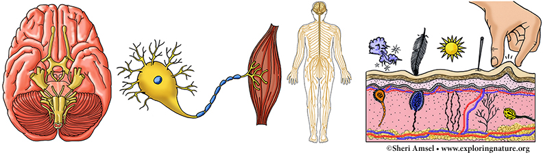
The nervous system is made up of the brain, spinal cord and all the nerves that run between your brain and spinal cord. Even though they are all connected, for convenience we break down the nervous system down into two parts – the central nervous system and the peripheral nervous system.
The Central Nervous System (CNS) is the brain and the spinal cord – which runs down your back inside the bony vertebral column. The CNS receives the information, decides what to do with it and gives the orders for action.
The Peripheral Nervous System (PNS) is all the nerves outside the cerebral nervous system. This includes the nerves coming off the spinal cord and the brain – the spinal nerves and the cranial nerves.
The nervous system controls how all the other body systems work in a huge communication network with messages coming in and going out every second. It keeps track of everything going on in the body and with the help of the endocrine system makes it run smoothly and in balance – it maintains homeostasis. Messages coming and going through the nervous system are communicated via electrical signals that are very quick and specific.
The nervous system has three jobs, which overlap to keep the body running smoothly.
1) It receives information (or stimulus) from outside and inside the body. Millions of tiny nerve cells – called sensory receptors, sense the stimuli. The stimulus is the sensory input. If you walk outside and see the sun shining, smell the flowers blooming, and hear the birds singing, you have gotten just three kinds of sensory input. Those are the three stimuli that are the most obvious that you notice right away though. Your brain is also getting sensory input from all kinds of body sensors all the time. You will know when you are hot, cold, hungry, thirsty, sleepy or sore. You can tell when you are lying down, standing up, falling, or get stung by a wasp Your brain gets the stimulus and tells you what it is.
2) It decides what to do with all that sensory input. This is called integration, because it integrates or brings together the stimulus with what you are going to do about it.
3) It triggers action. This is the motor response or motor output. Your muscles (or glands) respond to orders.
For example, while riding on a bike path, you come around a corner and see a dog in your way (sensory input). Your nervous system “integrates” the information (hitting the dog would hurt the dog and you). You slam on your brakes to stop the bike (motor response).
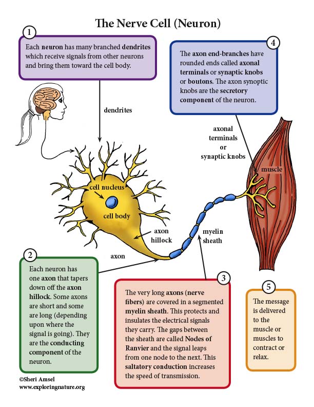
All those messages coming into and out of the brain are carried by nerve cells. Nerve cells (or neurons) carry nerve impulses – like an electrical cord carries an electrical signal. Nerve cells receive information through thousands of tiny finger-like projections called dendrites. They deliver all the information into the body of the nerve cell. They also have a very, very long tail called an axon. The axon carries the message to its destination. These axons are smaller than the tiniest thread, but travel together all over the body grouped together in bundles called tracts in the brain and spinal cord (CNS) and nerves in the rest of the body (PNS). You have thousands of nerves running signals all over your body all the time. The nerves branch into smaller and smaller pathways to pick up and drop off their messages to different parts of the body. The messages are then delivered to the muscles or glands that need to act.
Passing on the Message
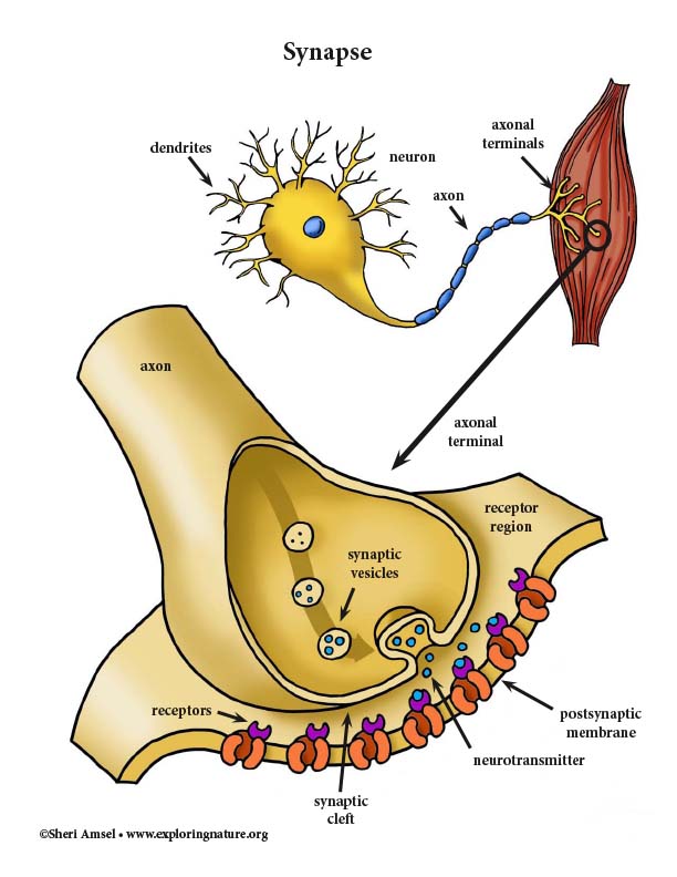
If a message has to go really far in the body (think about a strand thinner than a hair stretching between your brain and big toe) it sometimes takes two nerve cells to get it there. The first nerve cell will bring the message in through its dendrites and pass it to the next nerve cell. Then that nerve cell sends the message down its long axon to its final destination. The tiny space between the two nerve cells or the nerve cells and its target muscle is called a synapse. Instead of that electrical message jumping across this little gap or synapse, it triggers a chemical to squirt across and deliver the final message. This happens very fast. The chemical messenger is called a neurotransmitter.
A synapse is the junction between one neuron and another neuron or one neuron and an effector cell (muscle or gland cell). It is where the transfer of information occurs. A neuron may have up to 10,000 axonal terminals making contact through synapses. Synapses between two neurons have a presynaptic neuron and postsynaptic neuron. Most neuron to neuron synapses occur between the axonal endings of one and dendrites or cell body of the other. Synapses between a neuron and a muscles cell are called neuromuscular junctions. Synapses between a neuron and a gland cell are called neuroglandular junctions. Synapses can be electrical or chemical.
Electrical synapses provide low-resistance electrical pathways where ions flow from one neuron to the next. They are electronically coupled. These are called bridged junctions. This allows activity to happen all at once – to be synchronized. They are more common in human development. In adults, electrical synapses are most common in subconscious actions associated with cardiac and smooth muscle where rhythmic and sequential stimuli occurs.
Chemical synapses release and accept chemical neurotransmitters. They affect ion channels to either open or close which affects the permeability of membranes, which, in turn, determines membrane potential. At the site of a chemical synapse there is the axonal terminal of the transmitting neuron, which contains synaptic vesicles full of neurotransmitter molecules and the receptor region of the membrane. The space between them, though very small, is called the synaptic cleft filled with fluid. The nerve impulse moves down the axon as an electrical signal, but once it reaches the axonal terminal, it stimulates the release of neurotransmitter which crosses the synaptic cleft, binds with receptors on the receptor region and changes the membrane permeability to allow the stimulation of the postsynaptic cell.
The Brain
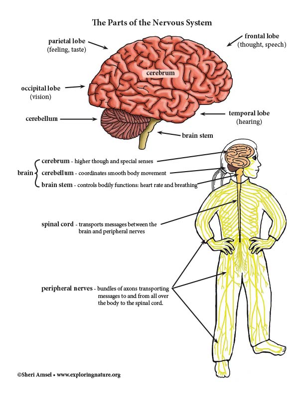
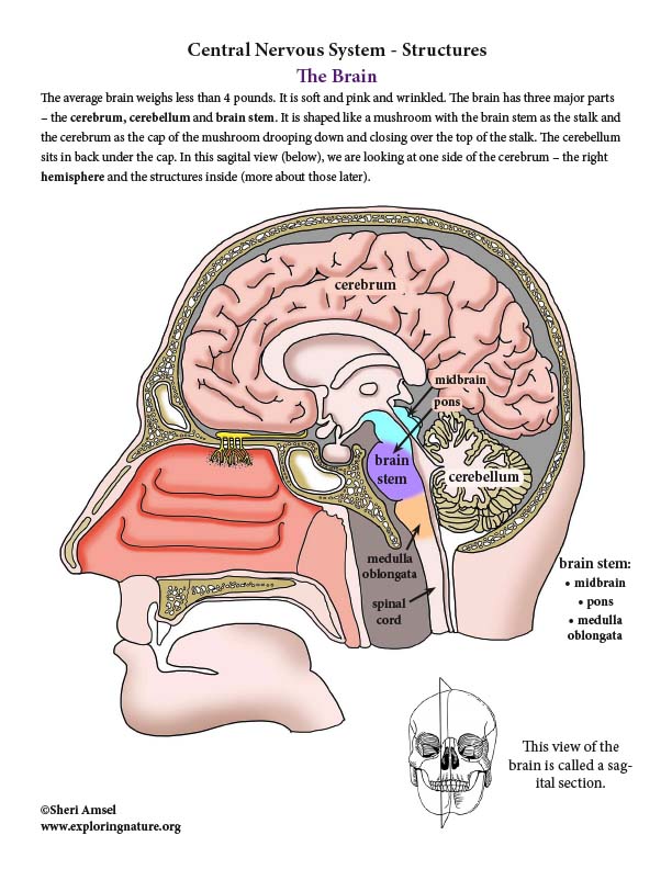
The average brain weighs less than 4 pounds. It is soft and pink and wrinkled. The brain has three major parts – the cerebrum, cerebellum and brain stem. It is shaped like a mushroom with the brain stem as the stalk and the cerebrum as the cap of the mushroom drooping down and closing over the top of the stalk. The cerebellum sits in back under the cap. In this sagital view (below), we are looking at one side of the cerebrum – the right hemisphere and the structures inside (more about those later).
The Cerebrum
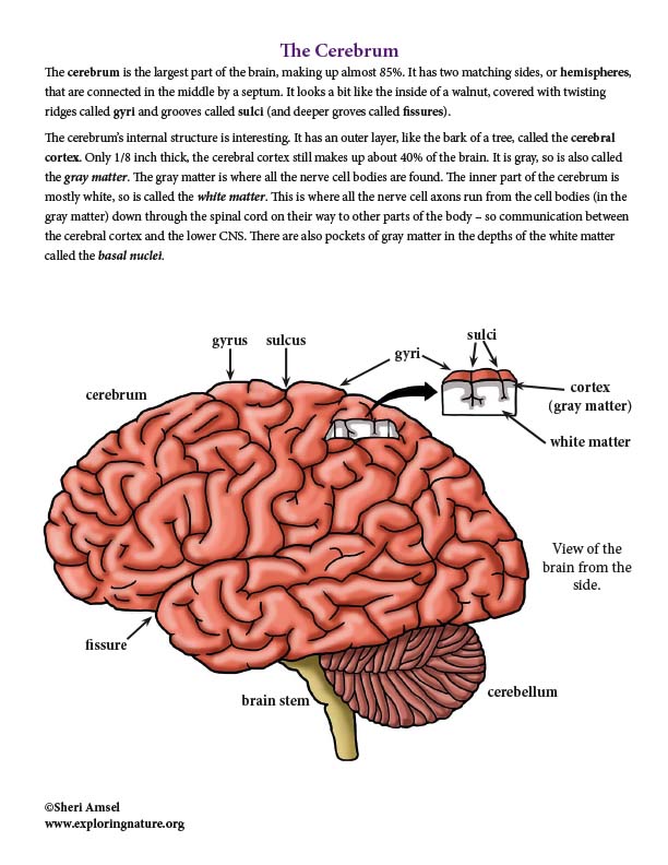
The cerebrum is the largest part of the brain, making up almost 85%. It has two matching sides, or hemispheres, that are connected in the middle by a septum. It looks a bit like the inside of a walnut, covered with twisting ridges called gyri and grooves called sulci (and deeper groves called fissures).
The cerebrum’s internal structure is interesting. It has an outer layer, like the bark of a tree, called the cerebral cortex. Only 1/8 inch thick, the cerebral cortex still makes up about 40% of the brain. It is gray, so is also called the gray matter. The gray matter is where all the nerve cell bodies are found. The inner part of the cerebrum is mostly white, so is called the white matter. This is where all the nerve cell axons run from the cell bodies (in the gray matter) down through the spinal cord on their way to other parts of the body – so communication between the cerebral cortex and the lower CNS. There are also pockets of gray matter in the depths of the white matter called the basal nuclei.
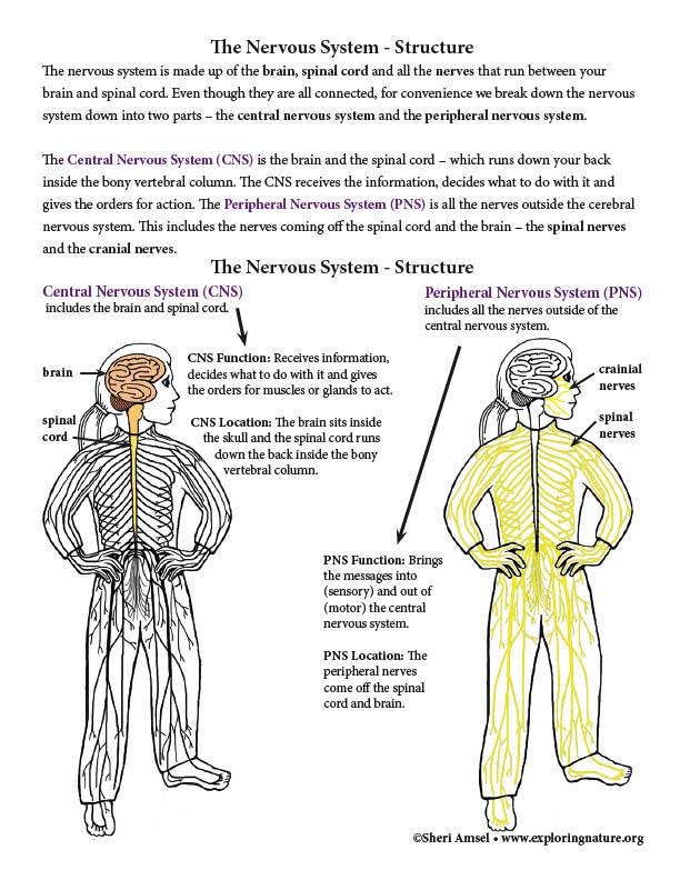
The Nervous System - Structure
The nervous system is made up of the brain, spinal cord and all the nerves that run between your brain and spinal cord. Even though they are all connected, for convenience we break down the nervous system down into two parts – the central nervous system and the peripheral nervous system.
The Central Nervous System (CNS) is the brain and the spinal cord – which runs down your back inside the bony vertebral column. The CNS receives the information, decides what to do with it and gives the orders for action. The Peripheral Nervous System (PNS) is all the nerves outside the cerebral nervous system. This includes the nerves coming off the spinal cord and the brain – the spinal nerves and the cranial nerves.
The Brain
The average brain weighs less than 4 pounds. It is soft and pink and wrinkled. The brain has three major parts – the cerebrum, cerebellum and brain stem. It is shaped like a mushroom with the brain stem as the stalk and the cerebrum as the cap of the mushroom drooping down and closing over the top of the stalk. The cerebellum sits in back under the cap. In this sagital view (below), we are looking at one side of the cerebrum – the right hemisphere and the structures inside (more about those later).
The Cerebral Cortex
The cerebral cortex is responsible for our consciousness, as such. It has:
1) Motor areas that control voluntary movement.
2) Sensory areas that make us aware of sensations (stimuli).
3) Areas that integrate all the sensory information coming in (vision, smell, hearing, taste, touch) with organized action to respond to it.
The cerebral cortex is where the human personality lives. It allows you talk, listen and understand what people are saying. It contains and organizes your memories – from what you had for breakfast to your memories of kindergarten. With it you understand the things around you, such as heat, cold, smells, and sights, and how you react to them. If you suddenly smell smoke, you might wonder for a spilt second before springing into action. You would jump up to see where it was coming from and if there was a fire to put out or escape. This is what your gray matter – your cerebral cortex, does for you.
It’s also important to note that:
1) No area of the cortex acts alone, so breaking down areas and function is a simplification.
2) The two cerebral hemispheres are control and feel the stimuli from the opposite side of the body. (This is why a left hemisphere stroke would affect the right side of the body.)
3) The two cerebral hemispheres are not exactly the same. Each side has specific areas unique to its hemisphere (e.g. speech center, etc.)
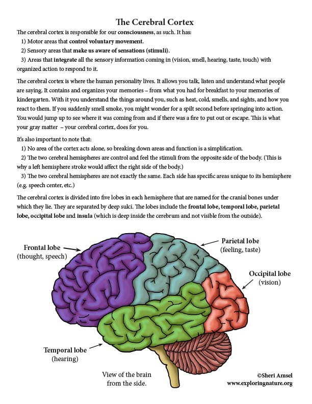
The cerebral cortex is divided into five lobes in each hemisphere that are named for the cranial bones under which they lie. They are separated by deep sulci. The lobes include the frontal lobe, temporal lobe, parietal lobe, occipital lobe and insula (which is deep inside the cerebrum and not visible from the outside).
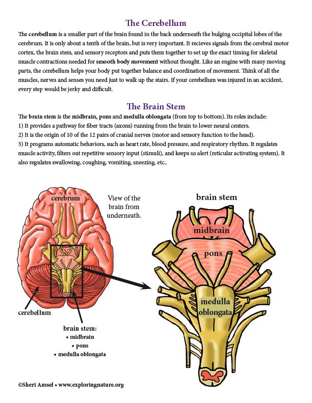
The Cerebellum
The cerebellum is a smaller part of the brain found in the back underneath the bulging occipital lobes of the cerebrum. It is only about a tenth of the brain, but is very important. It recieves signals from the cerebral motor cortex, the brain stem, and sensory receptors and puts them together to set up the exact timing for skeletal muscle contractions needed for smooth body movement without thought. Like an engine with many moving parts, the cerebellum helps your body put together balance and coordination of movement. Think of all the muscles, nerves and senses you need just to walk up the stairs. If your cerebellum was injured in an accident, every step would be jerky and difficult.
The Brain Stem
The brain stem is the midbrain, pons and medulla oblongata (from top to bottom). Its roles include:
1) It provides a pathway for fiber tracts (axons) running from the brain to lower neural centers.
2) It is the origin of 10 of the 12 pairs of cranial nerves (motor and sensory function to the head).
3) It programs automatic behaviors, such as heart rate, blood pressure, and respiratory rhythm. It regulates muscle activity, filters out repetitive sensory input (stimuli), and keeps us alert (reticular activating system). It also regulates swallowing, coughing, vomiting, sneezing, etc..
The Spinal Cord
The spinal cord runs from the brain down the back inside the protective vertebral column. It is made up of the very long, thin axons (nerve fibers) of thousands of nerve cells bringing their messages to and from the brain and body. It is also the site of spinal reflexes starting and ending in the spinal cord with no input from the brain.
The spinal cord is protected by the vertebral column and a tough layer of connective tissue meninges called the spinal dural sheath. Between the layers of the meninges is a fluid layer of cerebrospinal fluid that further protects the cord. The spinal cord tapers down at the first lumbar vertebra into the conus medularis.
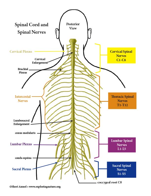
There are 31 pairs of spinal nerves that come off the spinal cord in paired roots. They leave the vertebral column through small holes – intervertebral foramina to reach the rest of the body. Spinal nerves are named by where they come off the spinal cord. There are 8 pairs of cervical spinal nerves, 12 pairs of thoracic nerves, 5 pairs of lumbar nerves, 5 pairs of sacral nerves and 1 pair of coccygeal nerves. The spinal cord ends at L1. The nerves to the lumbar and sacral region travel down, floating in cerebrospinal fluid, to reach their exits. Here they are called the cauda equina (horse’s tail).
The Spinal Nerves
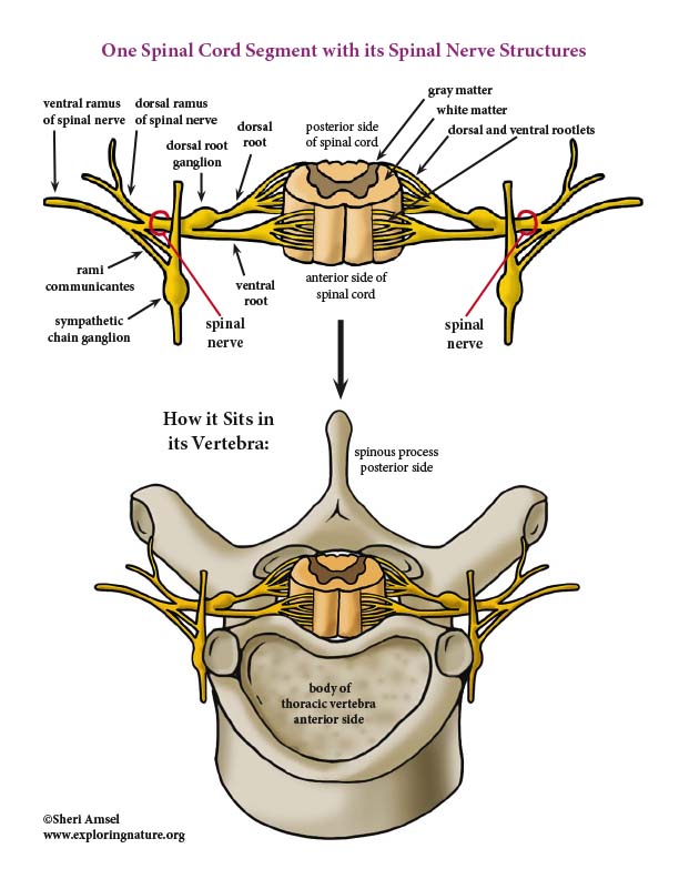
The 31 pairs of spinal nerves coming off the spinal cord are each made up of thousands of nerve fibers. These are all mixed nerves (both receiving and send sending signals), connected to the spinal cord by two roots – the doral root and ventral root. Each root is made up of rootlets that attach along that segment of the spinal cord which that spinal nerve serves.
The ventral root (anterior in the cord) contains the motor (efferent) messages coming out of the spinal cord. These fibers come off the anterior horn of the spinal cord gray matter, where motor neurons innervate the skeletal muscles.
The dorsal root (posterior side of the cord) contains the sensory (afferent) fibers coming into the spinal cord from sensory neurons in the dorsal root ganglia. They are bringing in sensory impulses from receptors in the body.
These spinal roots come together into the spinal nerves which exit the vertebral column on either side through small holes between the verterbra (intervertebral foramina), to reach the rest of the body. As they leave the foramen, each spinal nerve splits into a ventral ramus and a smaller dorsal ramus. They are mixed – containing both sensory and motor fibers. There is also, in the thoracic region, tiny rami communicantes coming off the ventral rami and containing automonic nerve fibers to regulate the body’s viscera.
The dorsal rami supply the posterior body trunk. The ventral rami supply the rest of the body trunk and the limbs (which is why they are bigger).
*Hint: Remember that dorsal and ventral roots are still inside the veterbral column and carry either sensory OR motor fibers, whereas dorsal and ventral rami are outside the vertebral column and are mixed, carrying both sensory and motor fibers in them. See the diagram below to clarify.
The Peripheral Nervous System
The peripheral nervous system includes nervous system structures outside the brain and spinal cord:
1) all peripheral nerves
2) sensory receptors and their associated ganglia
3) the efferent motor endings
The peripheral nervous system links the rest of your body to the spinal cord and brain.
1) It brings information in as sensory input via afferent sensory receptors. The nerves deliver the sensory input to the central nervous system (the spinal cord and brain) from all over your body – your skin, muscles, joints and even your internal organs. e.g. sunburn, pulled muscle, aching joints, even feeling full after a big meal.
2) It carry signals out as motor output via efferent motor endings. If the brain wants to tell the muscles to move, it sends the signal out through the peripheral nerves to those muscles and, in a split second, they move.
Sensory Receptors
Sensory Receptors sense changes in their environment. These changes are felt as stimuli. When a sensory receptor senses stimuli, it is activated and a nerve impulse travels up the nerve fibers (afferent fibers) to the CNS. Sensation and perception happen in the brain. The awareness of the stimulus is the sensation. The interpretation of the meaning of the stimulus is the perception. Sensory receptors are classified by their location, what they sense, and their structure.
Sensory Receptors by Location:
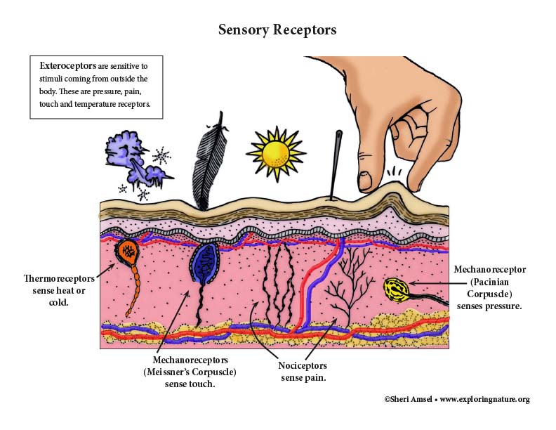
1) Exteroceptors are sensitive to stimuli coming from outside the body. These are pressure, pain, touch and temperature receptors and the special sense organs.
2) Interoceptors are sensitive to stimuli coming from inside the body. They respond to stimuli such as changes in body temperature, chemical changes, stretching of tissues.
3) Proprioceptors are sensitive to stimuli coming from inside the body – specifically the skeletal muscles, joints, tendons, ligaments and connective tissues associated with muscles and bones. They let us know where our body is in space. This allows you to move your body through the world without knocking into things.
Sensory Receptors by Stimulus Type:
1) Mechanoreceptors feel the stimulus of touch, pressure, stretch and vibrations.
2) Thermoreceptors feel temperature changes.
3) Chemoreceptors respond to chemical changes. This can be changes in blood chemistry, smell or taste.
4) Nociceptors feel stimuli that can damage the body as pain, e.g. excessive pressure, heat or cold, etc.
Sensory Receptors by Structure:
1) Simple sensory receptors are simple and found in the skin, muscles, mucous membranes, etc.
2) Complex sensory receptors are associated sense organs vision, smell, taste, hearing.
Spinal Reflexes
Sometimes messages coming into the nervous system need a very quick response. A reflex is a rapid, predictable motor response to a stimulus. It is involuntary. You do not have to learn how to do it or think about it. It is a message that doesn’t reach your brain before you act. Think about how fast you move when you touch a hot stove. The stimuli flies through the spinal cord and back out to give a quicker motor response. This is called a spinal reflex or a simple reflex arc. This is an important response to an emergency moment when your body needs to respond quicker than the brain can act.
Some reflexes do develop from practice. They are acquired reflex responses. They include situations where you can react quickly because of repetitive practice. Some examples of this are handling a sailboat in high winds, driving a car, and even playing tennis.
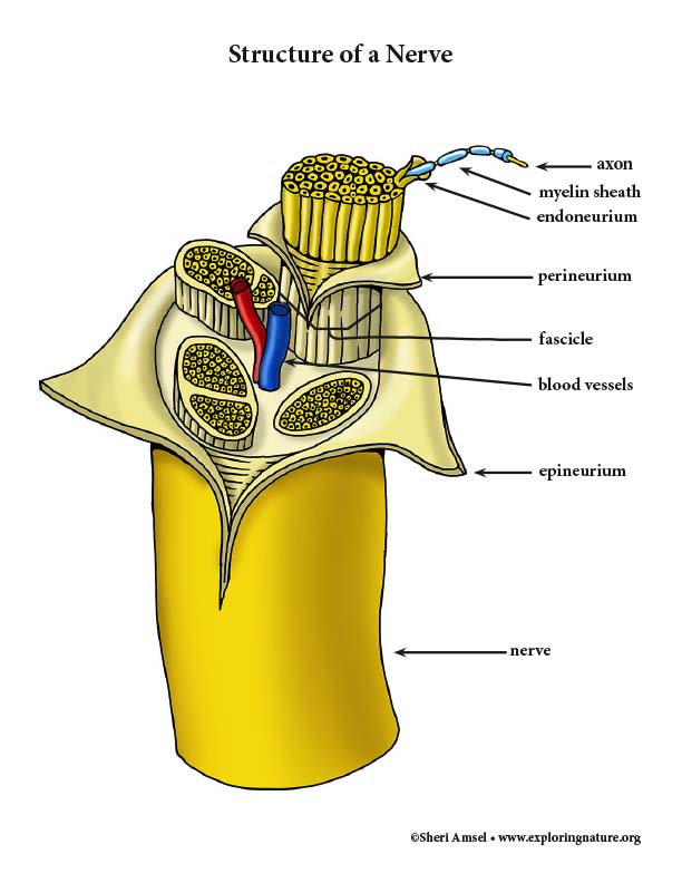
The Nerves
Nerves are made up of bundles of nerve cell axons in the peripheral nervous system (outside the brain and spinal cord) that are wrapped in protective connective tissue. Some of the axon are wrapped in myelin fibers and some are not. They are then covered in an endoneurium and bound into bundles called fascicles by perineurium. All the fascicles are bound in a protective fibrous sheath called an epineurium. All these together make up a nerve. Within all the wrapping of every nerve there are also blood vessels and lymphatic vessels.
Nerves are classified as cranial nerves (coming off the brain) or peripheral nerves (coming of the spinal cord). Some nerves carry only sensory (afferent) fibers, bringing impulses toward the CNS, so are called sensory nerves. Some nerves carry only motor (efferent) fibers carrying impulses away from the CNS, so are called motor nerves. Most nerves carry both sensory toward the CNS and motor fibers away from the CNS, so are called mixed nerves. Most nerves are mixed nerves.
Related Visual Aids:
Color Posters and Diagrams
Related Activities:
Labeling Pages
When you research information you must cite the reference. Citing for websites is different from citing from books, magazines and periodicals. The style of citing shown here is from the MLA Style Citations (Modern Language Association).
When citing a WEBSITE the general format is as follows.
Author Last Name, First Name(s). "Title: Subtitle of Part of Web Page, if appropriate." Title: Subtitle: Section of Page if appropriate. Sponsoring/Publishing Agency, If Given. Additional significant descriptive information. Date of Electronic Publication or other Date, such as Last Updated. Day Month Year of access < URL >.
Amsel, Sheri. "Nervous System Structures and Function (Advanced)" Exploring Nature Educational Resource ©2005-2024. December 13, 2024
< http://www.exploringnature.org/db/view/Nervous-System-Structures-and-Function-Advanced >