There are many different kinds of joints in the human body. The most common type are synovial joints.
Synovial joints have certain features unique to them:
1) hyaline cartilage covered articulating surfaces on the bones (to reduce friction)
2) fibrous articulating capsule that protects the joint (fluid-filled)
3) a joint cavity (fluid-filled)
4) synovial fluid to lubricate the joints
5) ligaments that reinforce the joint
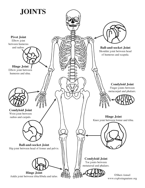
Examples of synovial joints include:
plane joints, hinge joints, pivot joints, condyloid joints, saddle joints and ball-and-socket joints.
Joint movements include:
• flexion (bending at joint to bring the two articulating bones closer together)
• extension (reverse of flexion), abduction (moving the limb away from the body)
• adduction (moving the limb back toward the body)
• circumduction (moving the limb in a cone shape)
• rotation (turning a limb on its own axis)
• pronation and supination, (radius and ulna)
• inversion and eversion (foot)
• protraction and retraction (jaw)
• elevation and depression (scapula)
• opposition (thumb)
Ball-and-socket joints are free moving synovial joints. Because they are free moving, they are unstable joints. Examples of ball-and-socket joints are the shoulder and hip.
With the shoulder joint (also called the glenohumeral joint), there are ligaments that reinforce it anteriorly. Tendons of four muscles encircle and fuse the joint to stabilize it. They are called the rotator cuff. Rotator cuff injuries are common in pitchers. The lack of tendons reinforcing the shoulder joint inferiorly allows free movement but also allows shoulder dislocation. The shoulder joint has many bursae – sacs of synovial fluid that lubricate where tendons, ligaments and muscle rubs against the bone.
With the hip joint there is not as much range of motion as the shoulder. The acetabular labrum is a fibrocartilage ring that depends the acetabulum of the pelvis in which the head of the femur sits to help stabilize it. Many ligaments reinforce the hip.
Hinge joints permit flexion and extension only. They have limited movement but a lot of stability. Examples of hinge joint are the elbow, knee and ankle.
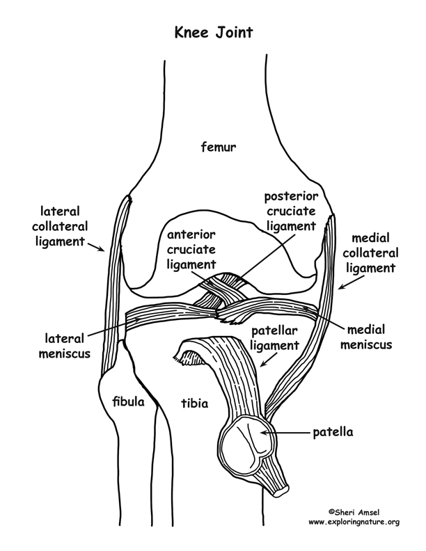
The knee joint (also called the tibiofemoral joint) is the largest joint of the body. It allows extension, flexion and some limited rotation and is reinforced by many ligaments.
Knee joint ligaments include:
1) The medial collateral ligament, which prevents lateral or medial angular movement of the knee joint. Its origin is the medial epicondyle of the femur. Its insertion is the medial tibial condyle and shaft.
2) The lateral collateral ligament, which prevents lateral or medial angular movement of the knee joint. Its origin is the lateral epicondyle of the femur. Its insertion is the head of the fibula.
3) The anterior and posterior cruciate ligaments cross each other and secure knee joint articulations. The anterior cruciate prevents over-extension of the knee joint. The posterior cruciate prevents over-flexion of the knee joint.
4) The patellar ligament extends from the patella to the tibial tuberosity (and is a continuation of the quadriceps muscle).
5) The medical meniscus and lateral meniscus are cartilage cups on the proximal end of the tibia in which the distal end of the femur sits. It helps stabilize the femur in articulation.
Despite all these ligaments, the knee joint is weight bearing and is the joint most susceptible to injury. The injury called “clipping” results from a blow to the side of the knee and involves injuries to the medial collateral ligament, medial meniscus and anterior cruciate ligament.
Other synovial joint types include:
Plane joints – which include the joints between the carpals, tarsals and vertebrae.
Pivot joints are when one bone rotates inside another. An example is the atlas and axis (cervical vertebrae) when you rotate your head in a “no” motion.
Condyloid joints have oval ends fitting into concave oval cups as with the knuckle joint.
Saddle joints are like condyloid joints but with more movement like the thumb joint.
*Test your knowledge of synovial joints in the labeling page included in the PDF below.