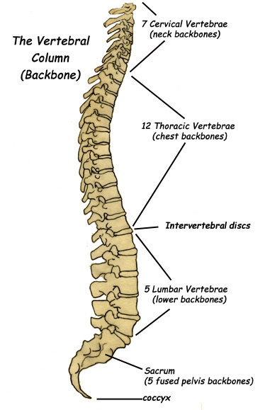
The vertebral column includes; 7 neck vertebra called cervical vertebra, 12 chest vertebra called thoracic vertebra, 5 lower back vertebra called lumbar vertebra, the sacrum (which makes up the back side of the pelvis) and the tail bone or coccyx.
The vertebral column is made up of 26 irregular bones. It is attached on top to the skull, which it holds up and to the pelvis on the bottom, where it is anchored to the lower limbs. It surrounds and protects the spinal cord inside it. It anchors the ribcage and the strong muscles of the back that hold the body upright. Each vertebra is separated by a cushion-like disc that helps pad the moving backbones as we move. The vertebral column is about 28 inches long in an average adult. It has an S-shape that helps it be strong and flexible.
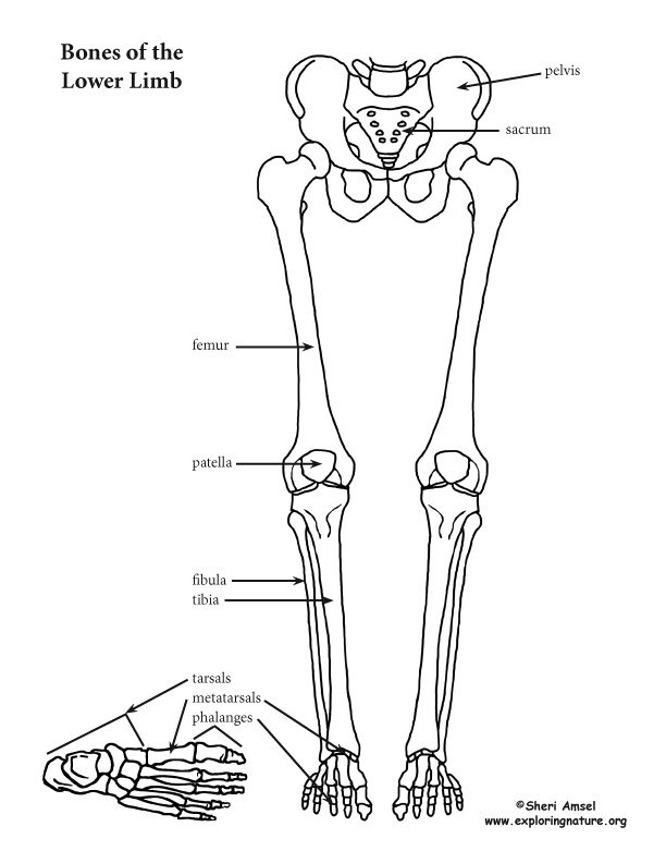
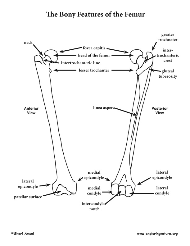
The lower limb includes the femur (thigh), the tibia (shinbone) and fibula (leg), and the bones of the foot: tarsals (ankle), metatarsals (foot) and phalanges (toes). The kneecap (patella) sits in front of the knee joint, inside a muscle tendon. The femur is the largest bone in the body and makes up about 1/4th of a person's height. It forms a ball and socket joint at the hip with the pelvis and a hinge joint at the knee with the tibia.
The hip joint is very important for leg movement and is supported by strong muscles and ligaments. Though it is a ball and socket joint like the shoulder, it is more stable and less moveable than that joint. The knee, a hinge joint, has less flexibility than the hip but is more stable. However, because the knee carries the bulk of the body's weight, it is often injured. The entire weight of the body sits on the foot. The foot acts as a lever to move the body forward when we walk or run.
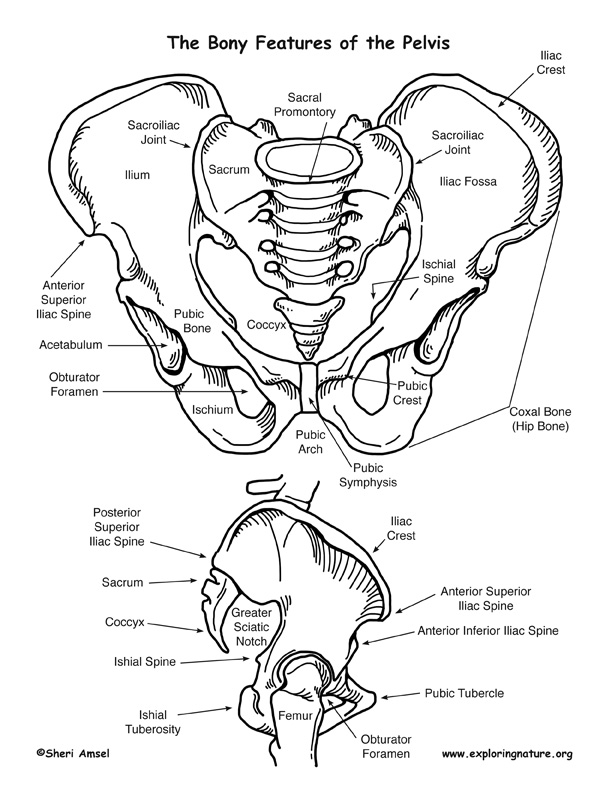
The pelvis, or pelvic girdle is formed by 2 hip bones (coxal bones). They come together in front and in back, they attach to the sacrum of the backbone. Each coxal bone is actually made up of 3 fused bones (ilium, ischium and pubis). On the outside of each coxal bone is a deep socket, called the acetabulum, where the head the leg bones (the femur) sits.
The pelvis supports and protects internal organs, attaches the lower limb to the body and with the lower limb supports the weight of the whole upper body. The hip joint is very important for leg movement and is supported by strong muscles and ligaments. Though it is a ball and socket joint like the shoulder, it is more stable and less moveable than that joint.
The femur is the largest and strongest bone in the body. It is a long bone making up about one fourth of your height and is the attachment point for some powerful muscles. At the hip (proximally), the femur's ball-shaped head joins (articulates) with the pelvis in the acetabulum and is secured by a strong ligament (ligamentum teres) attached to the fovea captitis on the head. The head attaches out (laterally) to a short neck that then attaches to the vertical shaft of the femur. Because of the way the head of the femur attaches to the side of the shaft, the neck is the weakest point and most prone to a break. Where the neck meets the shaft of the femur you will find the greater trochanter on the outside (lateral) and the medial trochanter on the inside (medial) which are connected by the intertrochanteric line in front (anteriorly) and the intertrochanteric crest in the back (posteriorly). The trochanters are attachment points for the powerful muscles of the thigh and butt. More muscle attachment sites are found on the back side of the femur (posteriorly) – the gluteal tuberosity which leads down the shaft to the ridge-like linea aspera. At the bottom (distally), the femur spreads into a wide base with the medial and lateral condyles joining (articulating) with the tibia. Between the condyles is the intercondylar notch. Outside of them are the lateral and medial epicondyles, which are attachment sites for more muscles. The patellar surface in the front (anteriorly) joins (articulates) with the kneecap (patella).
LS1.A: Structure and Function
• All living things are made up of cells, which is the smallest unit that can be said to be alive. An organism may consist of one single cell (unicellular) or many different numbers and types of cells (multicellular). (MS-LS1-1)
• Within cells, special structures are responsible for particular functions, and the cell membrane forms the boundary that controls what enters and leaves the cell. (MS-LS1-2)
• In multicellular organisms, the body is a system of multiple interacting subsystems. These subsystems are groups of cells that work together to form tissues and organs that are specialized for particular body functions. (MS-LS1-3)
When you research information you must cite the reference. Citing for websites is different from citing from books, magazines and periodicals. The style of citing shown here is from the MLA Style Citations (Modern Language Association).
When citing a WEBSITE the general format is as follows.
Author Last Name, First Name(s). "Title: Subtitle of Part of Web Page, if appropriate." Title: Subtitle: Section of Page if appropriate. Sponsoring/Publishing Agency, If Given. Additional significant descriptive information. Date of Electronic Publication or other Date, such as Last Updated. Day Month Year of access < URL >.
Amsel, Sheri. "Back, Pelvis and Lower Limb" Exploring Nature Educational Resource ©2005-2024. December 14, 2024
< http://www.exploringnature.org/db/view/Back-Pelvis-and-Lower-Limb >