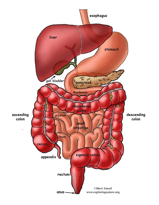
The digestive system takes in food, breaks it down (digests), sends the usable parts off into the blood and gets rid of the waste. There are many organs involved in the digestive system. They are broken down into two groups: The gastrointestinal tract (GI tract), which includes all the organs that food passes through in the body, and the accessory digestive organs, that break down food through action (like chewing) or chemicals (like saliva). The GI tract includes: the mouth, pharynx, esophagus, stomach, small intestine, and large intestine. The accessory digestive organs include: the teeth, tongue, salivary glands, liver, pancreas and gall bladder.
The mouth (oral cavity) is where digestion begins. In the mouth are the teeth, tongue and the salivary glands. The teeth chew the food and begin mechanical digestion. The tongue moves the food around to help break it down and mix it with saliva. Saliva is secreted by the salivary glands to begin the chemical breakdown of starchy foods (like bread). Food is chewed and rolled into a bolus (lump) to be swallowed. When you swallow food, it passes through the pharynx (back of the throat) down into the esophagus. The esophagus is a long, muscular tube about 10 inches long (25 cm). Food passes down it to reach the stomach. The muscles of the esophagus contract to squeeze the food downward. This is called peristalsis. Food must pass through the cardiac sphincter at the bottom end of the esophagus to enter the stomach. This muscular valve keeps the stomach contents, once mixed with stomach acids, from re-entering the esophagus and burning its unprotected lining.
The stomach is a storage tank for digesting food. Its walls contain a layer of muscle that can stretch if a lot of food is eaten. It can hold up to a gallon of food. When the stomach is empty, it shrinks back down and its walls fold up into wrinkles called rugae.
The lining of the stomach has lots of goblet cells that make and secrete a slimy mucous to protect the stomach lining from the powerful acids used to digest food. There are also gastric pits that make the gastric juice. Gastric juice is a mixture of the chemicals that digest food. It includes; hydrochloric acid (HCL) which kills bacteria on the foods you eat and helps the other chemicals work. Another chemical in gastric juice is pepsin, the chemical that digests proteins. The stomach is where protein digestion begins.
The stomach does both mechanical and chemical digestion. It churns the food and mixes it with the gastric juices. By the time it leaves the stomach, the food is broken down into a creamy paste called chyme. Now it is ready to move on to the small intestine. The pyloric sphincter is the muscular valve that regulated release of chyme into the small intestine.
The liver and the gall bladder are accessory organs of the small intestine. The liver is a large organ, weighing about 3 pounds. It has 4 lobes. The gall bladder is a tiny, green sac about 4 inches long (10 cm). The liver is one of the most important organs of the body. Only a small part of what it does has to do with digestion. It makes bile, which breaks down fat. It also takes the blood coming from the digestive tract and changes all the nutrients into forms the body can use, storing some. It cleans alcohol and drugs from the blood, stores vitamins and reuses the iron in old, worn out red blood cells. The gall bladder stores bile made in the liver.
The small intestine is about 6 feet long (2 meters) in an adult*, a hollow tube that twists and turns in a jumbled mass tucked inside the curve of the large intestine. It is divided into 3 parts: the duodenum, the jejunum and the ileum.
The small intestine is where digestion is completed and all the food nutrients are taken off (absorbed) into the blood. The digested food from the stomach (chyme) empties into the duodenum. It is here that the pancreas gland sends its pancreatic juice into the food. Also bile made in the liver and stored in the gall bladder enters here. Bile helps break down fats.
Inside the small intestine, the lining has tiny fingers called villi. Villi absorb the nutrients from the food passing through the small intestine. The nutrients then pass into the capillaries and are transported around the body to supply all our body systems.
Once all nutrients are extracted from the chyme, it is passed into the large intestine.
* If you could relax all the muscle of the small intestine you can stretch it out to about 20 feet long (6 meters).
The large intestine is about 4.5 feet long (1.5m) and wider than the small intestine. It is made up of the cecum, appendix, colon, rectum and anal canal. Food waste passes from the small intestine (ileum) into the cecum, then up the ascending colon, across the transverse colon, down the descending colon, through the sigmoid colon and into the rectum, where it is passed from the body through the anal canal.
The large intestine absorbs any left over water, vitamins and electrolytes (like sodium and chloride) in the food waste passing through it. The appendix does have some tissue that might help the immune system (lymphoid), but its tiny, twisted shape also traps bacteria, so often leads to serious imflammation, that can lead to death if not removed - appendicitis.
Testing:
Assess Digestive System comprehension with the Mutiple Choice Test.
Visual Aids and Assessment:
LS1.A Structure and Function
K-2 All organisms have external parts that they use to perform daily functions.
3-5 Organisms have both internal and external macroscopic structures that allow for growth, survival, behavior, and reproduction.
6-8 All living things are made up of cells. In organisms, cells work together to form
tissues and organs that are specialized for particular body functions.
9-12 Systems of specialized cells within organisms help perform essential functions of life. Any one system in an organism is made up of numerous parts. Feedback mechanisms maintain an organism’s internal conditions within certain limits and mediate behaviors.
When you research information you must cite the reference. Citing for websites is different from citing from books, magazines and periodicals. The style of citing shown here is from the MLA Style Citations (Modern Language Association).
When citing a WEBSITE the general format is as follows.
Author Last Name, First Name(s). "Title: Subtitle of Part of Web Page, if appropriate." Title: Subtitle: Section of Page if appropriate. Sponsoring/Publishing Agency, If Given. Additional significant descriptive information. Date of Electronic Publication or other Date, such as Last Updated. Day Month Year of access < URL >.
Amsel, Sheri. "Digestive System Overview" Exploring Nature Educational Resource ©2005-2024. December 13, 2024
< http://www.exploringnature.org/db/view/1052 >